
Table of Contents
Identification of bacteria using staining technique
Staining(Identification of bacteria)
It is a technique, in which we identified the bacteria by using different technique. In nature, two different types of bacteria are present and which cause different two disease, the bacteria are small in size and we cannot see them necked eye. So, with the help of microscope and a dye solution, bacteria are identified.
Staining is a technique by which we identify the nature, structure and features of bacteria by using different dye solution under microscope. For example, gram +ve and gram -ve bacteria is identify by gram staining with the help of dye.
Importance of staining

With the help of staining, it is very easy to see the bacteria and the size and shape of bacteria can be observed. It is also important in diagnosis of different infection disease.
Types of staining
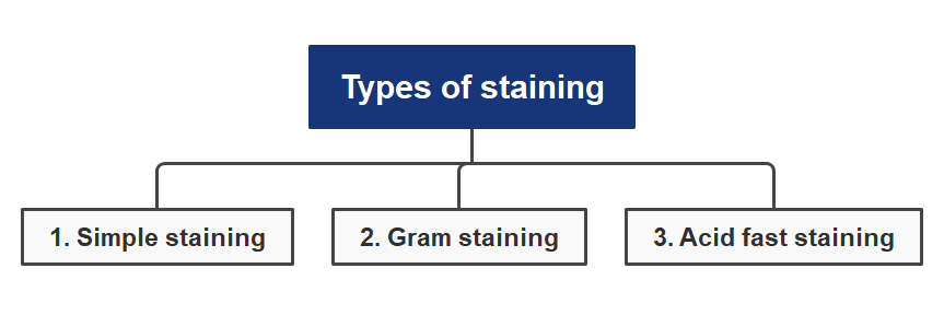
1. Simple staining
In this staining, we observe the morphological characteristic (shape and size) of bacteria. In which we use single stain dye such as crystal violet, safranine, methylene blue, malachite green.
Principle
Firstly, developed the negative charge on the surface of bacteria, so when we add positive charge dye on bacteria, it will attach easily (-ve attract +ve and attached easily).
When dye attached on the surface of bacteria, then bacteria will visible easily in also light background. Negative charge is developed on the surface of bacteria by releasing H+ ion or adding OH- ion.
Procedure
Firstly take – glass slide, cover slip, inoculation loop, culture media and microscope. Then, wash all these with ethanol solution after drying put under flaming for sterilization.
Now, take inoculation loop, streak into culture media in which bacteria attached on loop, then streak inoculation loop on glass side and add some drop of any indicator on surface of slide.
After that, allow slide for drying, then wash the slide under tap water for remove excess stain and wipe the below surface of slide with tissue paper.
Now, put the coverslip over the surface of slide when bacteria stained, then placed slide on surface of microscope. So, now see the bacteria in microscope and observe the bacteria.
Observation
The blue colored spherical shaped or rod-shaped bacteria is seen.
2. Gram staining
In this staining, we identified that bacteria are gram positive and gram negative. This staining technique was developed by Han’s Christian gram in 1884.
In this we use two or more dye at a time. In the basis of their structure, bacteria are classified into two categories: gram positive and gram negative.
Difference in gram positive and gram negative
| Gram positive staining | Gram negative staining |
| 1. In this peptidoglycan is present in multi-layer. | 1. In this peptidoglycan is present in single layer |
| 2. In this outer membrane is absent | 2. In this outer membrane is present |
| 3. In this periplasmic space is absent | 3. In this periplasmic space is present |
| 4. Cell wall 20-30nm thick & single layer | 4. Cell wall 8-12nm thick & two layers |
| 5. Example:- streptococcus, bacillus etc. | 5. Example:- Escherichia coli etc. |
1. In this peptidoglycan is present in multi-layer but in gram negative present in single layer.
2. Outer membrane is absent in gram positive but outer membrane is present in gram negative.
3. The periplasmic space is absent in gram positive but periplasmic space is present in gram negative.
4. The cell wall of gram positive is 20-30nm thick & single layer but in gram negative 8-12nm thick & two layers.
5. Example of gram positive is streptococcus and bacilli. Example of gram negative is Escherichia coli.
Procedure
Firstly take – glass slide, cover slip, inoculation loop, culture media and microscope. Then, wash all these with ethanol solution after drying put under flaming for sterilization.
Flame the inoculation loop and all the length of wire for complete sterilization. Now place the inoculation loop into the culture medium containing bacteria.
Now streak the inoculation loop on the surface of culture media and place the loop on glass slide.
Now add few drops of crystal violet indicator and allow to dry. Add grams in iodine solution, it acts as mordant which fix the crystal violet dye with cell wall.
Wash the slide with ethanol or acetone solution. Now add few drops of safranin indicator and allowed to dry.
Observation
If purple/blue color obtained then bacteria is gram positive because crystal violet not detached on washing, so safranin not attached.
If a red/pink color obtained then bacteria is gram negative because it detached crystal violet on washing with alcohol, so further when we apply safranin it attached with it give red/pink color.
3. Acid fast staining
It is used for those micro-organisms, which is wax like and impermeable cell wall (don’t have cell wall). These microorganisms do not identify by gram staining so used acid-fast staining because it doesn’t have cell wall.
Acid fast staining was developed by ziehl and identified by neelsen, so it is also called k/a ziehl neelsen staining.
Principle
Used to identified that bacteria are acid fast or non-acid fast organism. When we add Carbol-fuchsin dye as indicator, it reacts with inside material of bacteria in cytoplasm which is acidic and give a red color to bacteria. On washing with alcohol acid fast same red color but non-acid fast decolorized.
Procedure
Firstly take – glass slide, cover slip, inoculation loop, culture media (in which bacteria present), microscope (for see bacteria after staining). After that, wash all the with ethanol solution then allow for drying, after drying put all these under flaming for complete sterilization.
Now take inoculation loop, streak into culture media in which bacteria attached on loop then a streak inoculation of loop on glass slide and make smear on it.
Now add few Carbol-fuchsin dye (primary dye) on surface of bacteria and allow for dry, then wash it with alcohol.
Then further add some methylene blue (counter stain). Again, wash the slide with water, then dry and observe it by seeing it on microscope.
Observation
If bacteria give red/pink color it acid fast and if bacteria give blue color it non-acid fast.
Identification of bacteria using staining technique
Staining is a technique used to identify different types of bacteria. It involves applying dyes to the bacteria, which help distinguish them from other microorganisms. The dyes used in staining vary depending on the type of bacteria being identified. For example, Gram staining is often used for the identification of Gram-positive and Gram-negative bacteria. Staining techniques are also used to differentiate between species and strains of bacteria. Identification of bacteria using staining technique.

Staining techniques have been around for centuries and are still widely used today by medical professionals, researchers, and scientists alike. They provide an effective way to quickly and accurately identify different types of bacteria without having to perform complex laboratory tests or rely on expensive equipment. Staining can be a powerful tool in identifying bacterial infections as well as helping with diagnosis and treatment decisions. Identification of bacteria using staining technique.

Generally I don’t read post on blogs, but I would like to say that this write-up very forced me to try and do so! Your writing style has been amazed me. Thanks, quite nice article.
Hi mimprovement.com admin, Good work!